| Electrical Communication is a free textbook on the basics of communication technology. See the editorial for more information.... |

|

Home  Fundamentals of Acoustics Fundamentals of Acoustics  Hearing Hearing |
|||||


|
|||||
HearingThe ear consists (Fig. 14) of the three following parts:
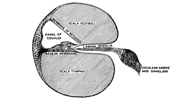
The cochlea is filled with fluid and is divided lengthwise into three parts by the basilar membrane and the membrane of Reissner. There are thus three parallel spiral canals, a cross section of which is shown in Fig. 15. The scala vestibuli terminates at one end on the oval window O, and the scala tympani terminates on the round window E. These two canals are connected at the extreme end of the spiral. The membrane of Reissner is very thin, and any impulses transmitted to the fluid in the scala vestibuli by the foot of the stirrup at the oval window are readily transmitted through this membrane to the fluid of the canal of cochlea. The flexible basilar membrane extends toward the upper end of the cochlea, dividing it as shown. An impulse transmitted to the fluid of the scala vestibuli will readily pass to the canal of cochlea and will set the basilar membrane in motion.
The organ of Corti containing the nerve terminals in the form of small hairs extending into the canal of cochlea is along one side of the basilar membrane. Lying over these small hairs is a soft, loose membrane called the tectorial membrane. The process of hearing is as follows.21 If a low-frequency note, below 20 cycles per second, impinges on the eardrum, this sound variation is transferred as a mechanical impulse to the fluid of the scala vestibuli. This fluid is in direct contact (at the far end) with that of the scala tympani, as previously mentioned. At this low frequency, the liquid offers little reactance, owing to its mass, and therefore the liquids in the two canals move bodily back and forth. The basilar membrane is not affected, and accordingly no sound sensation is produced. If a 1000-cycle tone is impressed on the ear, however, the mass reactance of the fluid is great enough so that the liquid does not move back and forth, and the impulse is transmitted through the membrane of Reissner to the canal of cochlea. The basilar membrane is caused to vibrate, and at some one spot the vibration will be greatest. The relative motion between the tectorial membrane and the basilar membrane then causes the small hairs to stimulate the nerve endings at their base, and this sends a sound sensation to the brain. If a tone of higher frequency is used, a different part of the basilar membrane will vibrate with greatest amplitude, and thus different nerve endings will respond. When the frequency reaches and exceeds about 20,000 cycles per second, the hammer, anvil, stirrup, and associated parts absorb most of the sound energy, and little is transmitted to the inner ear; thus, the upper limit of audition results. It can be shown21 that these three small bones together with the membranes on which they terminate act as a transformer to match the low impedance of the air to the high impedance of the fluid of the inner ear.
|
|||||
Home  Fundamentals of Acoustics Fundamentals of Acoustics  Hearing Hearing |
|||||
Last Update: 2011-04-05


