Lectures on Physics has been derived from Benjamin Crowell's Light and Matter series of free introductory textbooks on physics. See the editorial for more information....
Why would blue or violet light be the best for microscopy?
2
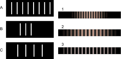
Match gratings A-C with the diffraction patterns 1-3 that they produce.
Explain.
3
The beam of a laser passes through a diffraction grating, fans out,
and illuminates a wall that is perpendicular to the original beam, lying at a
distance of 2.0 m from the grating. The beam is produced by a heliumneon
laser, and has a wavelength of 694.3 nm. The grating has 2000 lines
per centimeter. (a) What is the distance on the wall between the central
maximum and the maxima immediately to its right and left? (b) How
much does your answer change when you use the approximation
sin θ ≈ θ ?
√
4
When white light passes through a diffraction grating, what is the
smallest value of m for which the visible spectrum of order m overlaps the
next one, of order m+1? (The visible spectrum runs from about 400 nm to
about 700 nm.)
5
Ultrasound, i.e. sound waves with frequencies too high to be audible,
can be used for imaging fetuses in the womb or for breaking up kidney
stones so that they can be eliminated by the body. Consider the latter
application. Lenses can be built to focus sound waves, but because the
wavelength of the sound is not all that small compared to the diameter of
the lens, the sound will not be concentrated exactly at the geometrical
focal point. Instead, a diffraction pattern will be created with an intense
central spot surrounded by fainter rings. About 85% of the power is
concentrated within the central spot. The angle of the first minimum
(surrounding the central spot) is given by sin θ = 1.22 λ/b, where b is the
diameter of the lens. This is similar to the corresponding equation for a
single slit, but with a factor of 1.22 in front which arises from the circular
shape of the aperture. Let the distance from the lens to the patient's
kidney stone be L=20 cm. You will want f>20 kHz, so that the sound is
inaudible. Find values of b and f that would result in a usable design,
where the central spot is small enough to lie within a kidney stone 1 cm in
diameter.
6
For star images such as the ones in the photo in section 5.6, estimate
the angular width of the diffraction spot due to diffraction at the mouth of
the telescope. Assume a telescope with a diameter of 10 meters (the largest
currently in existence), and light with a wavelength in the middle of the
visible range. Compare with the actual angular size of a star of diameter
109 m seen from a distance of 1017 m. What does this tell you?
7
Under what circumstances could one get a mathematically undefined
result by solving the double-slit diffraction equation for θ? Give a physical
interpretation of what would actually be observed.
8
When ultrasound is used for medical imaging, the frequency may be as
high as 5-20 MHz. Another medical application of ultrasound is for
therapeutic heating of tissues inside the body; here, the frequency is
typically 1-3 MHz. What fundamental physical reasons could you suggest
for the use of higher frequencies for imaging?
9
The figure below shows two diffraction patterns, both made with the
same wavelength of red light. (a) What type of slits made the patterns? Is it
a single slit, double slits, or something else? Explain. (b) Compare the
dimensions of the slits used to make the top and bottom pattern. Give a
numerical ratio, and state which way the ratio is, i.e., which slit pattern
was the larger one. Explain.
10
The figure below shows two diffraction patterns. The top one was
made with yellow light, and the bottom one with red. Could the slits used
to make the two patterns have been the same?
11
The figure below shows three diffraction patterns. All were made
under identical conditions, except that a different set of double slits was
used for each one. The slits used to make the top pattern had a center-tocenter
separation d=0.50 mm, and each slit was w=0.04 mm wide.
(a) Determine d and w for the slits used to make the pattern in the
middle. (b) Do the same for the slits used to make the bottom pattern.
12
The figure shows a diffraction pattern made by a double slit, along
with an image of a meter stick to show the scale. The slits were 146 cm
away from the screen on which the diffraction pattern was projected. The
spacing of the slits was 0.050 mm. What was the wavelength of the light?
13
Sketch the diffraction pattern from the figure on your paper. Now
consider the four variables in the equation λ/d=sin θ/m. Which of these
are the same for all five fringes, and which are different for each fringe?
Which variable would you naturally use in order to label which fringe was
which? Label the fringes on your sketch using the values of that variable.

 Optics