| Basic Radio is a free introductory textbook on electronics based on tubes. See the editorial for more information.... |
|
Home  Electronic Devices Cathode-Ray Tubes Magnetic Focusing Electronic Devices Cathode-Ray Tubes Magnetic Focusing |
||||||||||


|
  |
|||||||||
|
Magnetic FocusingAuthor: J.B. Hoag
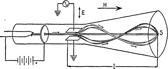
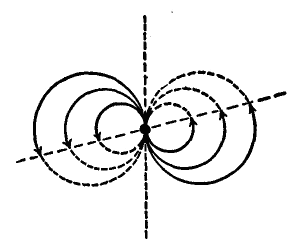
In Fig. 21 P, electrons from the hot filament are accelerated and collimated so as to focus at the center of the screen S. An alternating potential is applied across the deflection plates and sweeps the electron beam back and forth. A line instead of a spot is then observed on the fluorescent screen. Finally, a magnetic field, H, is applied, with its lines of force parallel to the axis of the tube and extending along the entire path of the electrons. This field does not change the forward motion of the electrons but it does act upon the transverse motion produced by the deflection plates. The electrons will be deflected in circular paths in planes at right angles to the axis of the tube at the same time that they move down the tube. The combination of their circular and forward motions is such that the electrons travel in helical paths down the tube, as shown in the figure. If it were possible to see the electrons from the screen end while they moved down the tube, it would be found that the circular paths of the various electrons are all tangent to the axis of the tube. This is indicated in Fig. 21 Q, where the black dot represents the intersection of the axis with the screen.
Fast electrons travel in larger circles, and vice versa, but the time of rotation is the same for all. (This is also the basic law of cyclotrons.)
By analogy, we may liken the magnetic field to a converging lens whose focal length is proportional to the velocity of the electrons and inversely proportional to the strength of the magnetic field. Thus, a strong magnetic field, twisting the electrons in the manner just described, is the equivalent of a lens. It is not necessary that the magnetic lens extend along the entire path of the electron beam in order that it shall serve as a converging lens. In fact, the magnetic field may be confined to an exceedingly-short distance along the axis of the tube and still serve to focus the electrons sharply on the screen.
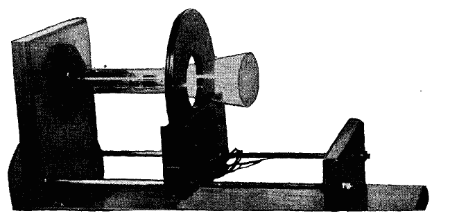
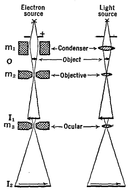
In Fig. 21 R, the thin disc-shaped magnetic field produces a magnetic field which extends along the axis of the cathode-ray tube only a few centimeters. With smaller diameter magnetic lenses, the field can be confined to only a few millimeters. Such " thin lenses " are used in electron microscopes. The principle of the electron microscope can be seen from Fig. 21 S.
Here a stream of high velocity electrons are rendered parallel by a magnet m1. They pass through a very thin slice of some object which is to be magnified. Some of the electrons are stopped by the object and others get through, just as some of the X-rays passing through the human body are stopped by the bones while others get through the flesh. Electrons which pass through various parts of the object o are focused by the magnetic lens m2 to form an image of the object at I1. This image is a shadowgraph, since it is produced by transmission through the object rather than by reflection from it.Furthermore, with m2 close to the object and I1 far away, the image is greatly magnified, as much so as can be accomplished with an ordinary optical microscope. Electrons which pass I1 are again focused by the third magnet m3 to the new and again greatly magnified image I2. A fluorescent screen or photographic plate is placed at I2. Magnifications as great as 100,000-fold can be produced by this double magnification system. But of still greater importance is the fact that the small parts of the object, when thus magnified, can be distinguished one from the other. Stated technically, the resolving power is much greater in the electron microscope than in the optical microscope. In fact, objects as small as 0.000,000,5 cm. have been observed and studied with this instrument. This is an extension of approximately 100 times that of optical instruments.
|
||||||||||
| Home Electronic Devices Cathode-Ray Tubes Magnetic Focusing |
|
|||||||||
Last Update: 2009-11-01